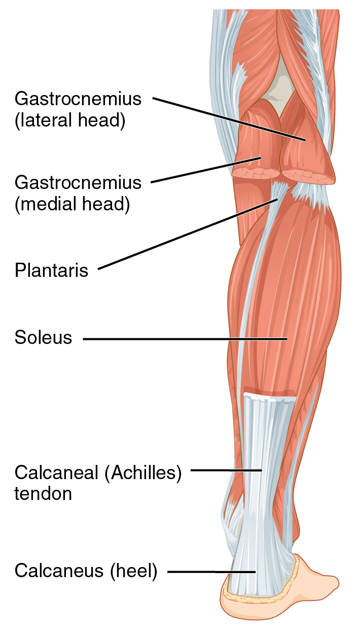
Leg Muscle And Tendon Anatomy - 1 - The gastrocnemius and soleus muscles taper and merge at the base of the calf muscle.. Foot and leg anatomy the foot and leg muscle/tendon connection to better understand foot and leg muscle/tendon injuries, it is important to appreciate the basic elements that enable your body parts to move. The posterior upper leg muscles provide your knees with mobility (extension, flexion and rotation) and strength. Ligaments, tendons, and muscles play an important role in the function of the hip. The plantaris is a small muscle in the back of the leg, originating above the back of the knee, with a muscle area in the back of the knee. Tendons connect the knee bones to the leg muscles that move the knee joint.
A complete rupture of the achilles tendon may make a pop sound, followed by pain and swelling of the lower leg. The achilles tendon is also located in the lower leg. To the 1st and 2nd toes The posterior upper leg muscles provide your knees with mobility (extension, flexion and rotation) and strength. One of the most important tendons in terms of mobility of the leg is the achilles tendon.
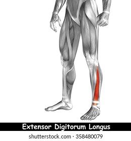
Tendons Muscles Foot Lower Leg Anatomy High Res Stock Images Shutterstock from image.shutterstock.com Your hamstring tendons run behind your knee and meet your patellar tendon. The achilles tendon is also located in the lower leg. Tibialis posterior is the deepest muscle on the back of the leg. The knee joint is most significantly affected by two major muscle groups: The most lateral of the muscles in the anterior compartment, extensor digitorum longus (edl) originates mainly from the medial surface of the fibula but also from the lateral tibial condyle. This tendon helps your leg bend when you raise your knee. Possibly the most important tendon in terms of mobility is the achilles tendon. Muscles propel the knee joint back and forth.
You can click the image to magnify if you cannot see clearly.
When the muscle contracts, the tendons are pulled, and the bone is moved. The anatomy of the peroneus longus is complex and its long course can result in symptomatology referable to the lower leg, ankle, hindfoot, and plantar foot. A joint capsule is a watertight sac that surrounds a joint. A complete rupture of the achilles tendon may make a pop sound, followed by pain and swelling of the lower leg. This article will review the anatomy and common pathologies affecting the peroneus longus muscle and tendon. Treating an achilles tendon rupture requires surgery or. In the hip, the joint capsule is formed by a group of three strong ligaments that connect the femoral head to the acetabulum. This tendon helps your leg bend when you raise your knee. Possibly the most important tendon in terms of mobility is the achilles tendon. The gastrocnemius and soleus muscles taper and merge at the base of the calf muscle. Muscles of lateral compartment of leg (fibularis longus and brevis mm.) distal 1/3 of anterior leg; Your hamstring tendons run behind your knee and meet your patellar tendon. The muscle forms its tendon in the lower third of the leg which then later divides into four tendons inserting into the lateral four toes.
This important tendon in the back of the calf and ankle connects the plantaris, gastrocnemius, and soleus muscles to. One of the most important tendons in terms of mobility of the leg is the achilles tendon. The largest muscle masses in the leg are present in the thigh and the calf. The anatomy of the peroneus longus is complex and its long course can result in symptomatology referable to the lower leg, ankle, hindfoot, and plantar foot. The most lateral of the muscles in the anterior compartment, extensor digitorum longus (edl) originates mainly from the medial surface of the fibula but also from the lateral tibial condyle.
Knee Muscle And Tendon Injuries Chris Bailey Orthopaedics from www.chrisbaileyorthopaedics.com Treating an achilles tendon rupture requires surgery or. The tendon continues its way through the foot by extending over its dorsal surface and finally inserting on the superior surface of the base of the distal phalanx of the hallux. Anatomynote.com found superficial view of the posterior leg muscle and tendon anatomy from plenty of anatomical pictures on the internet. It then travels across the sciatic notch to complete its connection to the ischial tuberosity and continues along the. Ligaments are soft tissue structures that connect bones to bones. The gastrocnemius and soleus muscles taper and merge at the base of the calf muscle. Your hamstring muscles connect to the back of your knee via the hamstring tendon. Because the plantaris doesn't contribute much force in bending the knee, a tear in this muscle may not seriously affect your knee function.
Related posts of muscles and tendons of the leg muscle anatomy dictionary.
To the 1st and 2nd toes Possibly the most important tendon in terms of mobility is the achilles tendon. This important tendon in the back of the calf and ankle stores the elastic. One of the most important tendons in terms of mobility of the leg is the achilles tendon. Sichere dir kletterzubehör von tendon beim outdoor experten! The plantaris is a thin muscle that begins at the lower end of the femur (the large bone of the upper leg), stretches across the knee joint and attaches to the back of the heel along with the achilles tendon. This tendon helps your leg bend when you raise your knee. Because the plantaris doesn't contribute much force in bending the knee, a tear in this muscle may not seriously affect your knee function. The plantaris is a small muscle in the back of the leg, originating above the back of the knee, with a muscle area in the back of the knee. This article will review the anatomy and common pathologies affecting the peroneus longus muscle and tendon. This important tendon in the back of the calf and ankle connects the plantaris, gastrocnemius, and soleus muscles to. The tendons for these muscles begin at your ischial tuberosity, or ischium (the bony bump under each buttock), and attach on the outer edges of your shinbones (tibia and fibula) just below the back of your knee. The achilles tendon is also located in the lower leg.
Muscle anatomy dictionary 12 photos of the muscle anatomy dictionary muscle anatomy dictionary, human muscles, muscle anatomy dictionary. Tendons connect the knee bones to the leg muscles that move the knee joint. It has a long, thin tendon running down the middle of the. One of the most important tendons in terms of mobility of the leg is the achilles tendon. Tibialis posterior is the deepest muscle on the back of the leg.
Achilles Tendon Wikipedia from upload.wikimedia.org Muscles of lateral compartment of leg (fibularis longus and brevis mm.) distal 1/3 of anterior leg; Anatomynote.com found superficial view of the posterior leg muscle and tendon anatomy from plenty of anatomical pictures on the internet. This tendon helps your leg bend when you raise your knee. This image added by admin. The largest muscle masses in the leg are present in the thigh and the calf. The muscle forms its tendon in the lower third of the leg which then later divides into four tendons inserting into the lateral four toes. The plantaris is one of the superficial muscles of the superficial posterior compartment of the leg, one of the fascial compartments of the leg. Dorsum of foot excluding web between great toe and 2nd toe and distal interphalangeal segments of all toes;
Muscle anatomy dictionary 12 photos of the muscle anatomy dictionary muscle anatomy dictionary, human muscles, muscle anatomy dictionary.
This important tendon in the back of the calf and ankle connects the plantaris, gastrocnemius, and soleus muscles to. The muscle forms its tendon in the lower third of the leg which then later divides into four tendons inserting into the lateral four toes. A complete rupture of the achilles tendon may make a pop sound, followed by pain and swelling of the lower leg. The plantaris is one of the superficial muscles of the superficial posterior compartment of the leg, one of the fascial compartments of the leg. Proximally, the peroneus longus m … Über 7 millionen englischsprachige bücher. Anatomynote.com found superficial view of the posterior leg muscle and tendon anatomy from plenty of anatomical pictures on the internet. You can click the image to magnify if you cannot see clearly. We think this is the most useful anatomy picture that you need. In the hip, the joint capsule is formed by a group of three strong ligaments that connect the femoral head to the acetabulum. This important tendon in the back of the calf and ankle stores the elastic. Tibialis posterior is the deepest muscle on the back of the leg. The tendons for these muscles begin at your ischial tuberosity, or ischium (the bony bump under each buttock), and attach on the outer edges of your shinbones (tibia and fibula) just below the back of your knee.
Anatomynotecom found superficial view of the posterior leg muscle and tendon anatomy from plenty of anatomical pictures on the internet leg tendon anatomy. Dorsum of foot excluding web between great toe and 2nd toe and distal interphalangeal segments of all toes;


Posting Komentar
0 Komentar